What is precision PCI?
Precision PCI is a term used to emphasise the importance of evidence-based treatment (stenting) strategy that is guided by coronary physiology (Is this narrowing truly restricting blood flow?) and lesion morphology (Is this narrowing dangerous and at risk of causing heart attack?)
Why do we need coronary physiology and intracoronary imaging?
Coronary angiography, just like clinical medicine, is part-art and part-science. Taking x-ray pictures of the arteries from different angles will allow some appreciation of the severity of the narrowing; but sometimes it can be very difficult to be certain whether a specific narrowing requires or will benefit from a stent.
To quantitatively evaluate the severity of specific narrowing, we can use a pressure-sensitive wire to measure the pressure drop across the narrowing - this will then allow us to infer the significance of blood flow obstruction. By doing so, pressure-wire technology allow us to be guided by science whether you will require a stent for a specific narrowing - hence the term precision PCI.
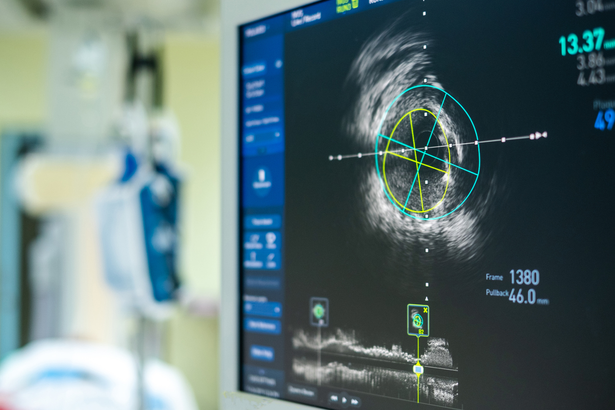
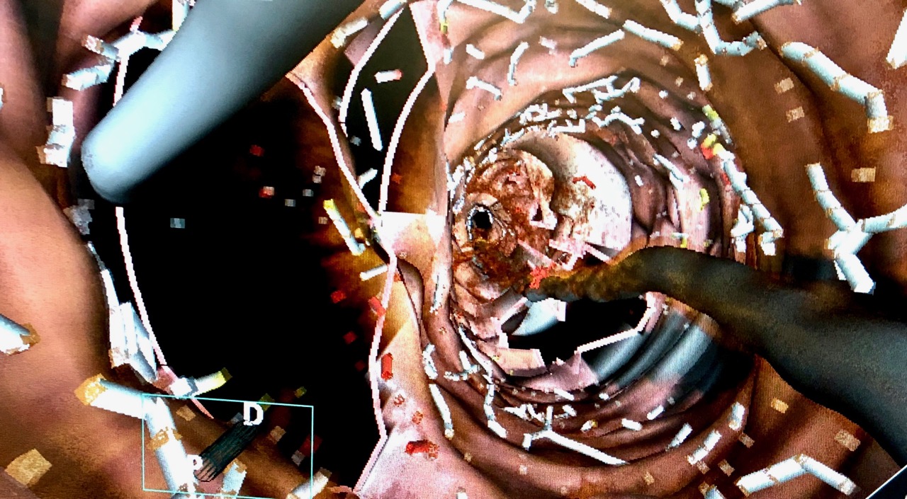
To allow for even more precise stenting, Dr Chai often deploys state-of-the-art intracoronary imaging techniques, such as co-registered optical coherence tomography (OCT) or intravascular ultrasound (IVUS) to examine the composition of the blood vessel underlying the narrowing as well as the surface anatomy at the "landing zone" either side of the narrowed segment to ensure optimal stenting result.
Precision PCI is particularly beneficial in patients with calcified heart arteries or patients with diabetic related coronary artery disease. These adjunctive diagnostic techniques are often performed as part of the treatment package to ensure the most long-lasting outcome.