Common presenting symptoms:
Unexplained (cryptogenic) stroke/mini stroke; breathlessness; swelling; detection of "murmur"
Overview:
Every one of us was born with a small “hole in the heart”. This small hole is a necessity for foetal life within the maternal circulation. It allows maternal (oxygenated) blood to bypass the foetal lung (which would be filled with amniotic fluid and practically useless for gas exchange inside the womb), to reach the foetal left heart in order to be pumped around to supply the foetal organs and development.
At birth, the small flap of tissue will usually close off this small hole (foramen ovale) when the newborn starts to inflate the lung. It usually fuses permanently at infancy. However, about 25% of the population this flap fails to shut close and has a residual patent foramen ovale (PFO). The rationale is that with a small shunt between the left and right heart, sudden reversal of pressure gradient may result in small blood clots to cross from right to left heart resulting in a stroke or mini-stroke. This is particularly important in patients who do professional or recreational diving / perform exercises with straining and Valsalva manoeuvres. Clearly, not 25% of the population will suffer a stroke or mini-stroke, therefore mechanical closure of PFO remains a little controversial and careful selection of cases is absolutely crucial.
In some cases, the residual hole (shunt) is considered so large that a septal defect is diagnosed. In such case, there could be haemodynamic effect caused by the shunt with chamber dilatation and sometimes even symptom of heart failure.
How do we investigate?
Often referred by stroke phyicians, if there is no alternative explanation of a stroke or possible stroke, we must entertain the idea of the presence of a PFO / shunt. In such case, our cardiologists will recommend one or more potential investigative tests such as:-
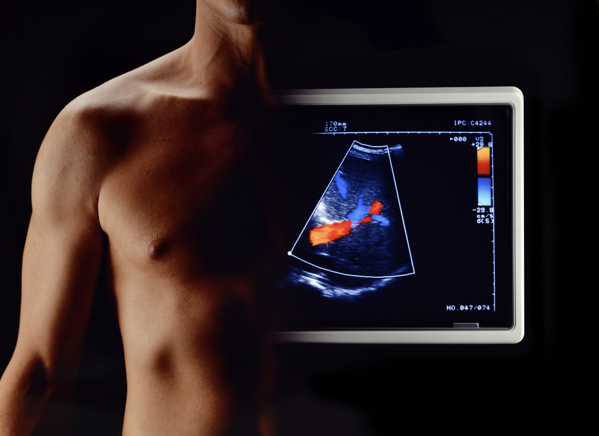
- Echocardiography to assess any abnormal shunt between left / right heart chambers
- Bubble contrast echocardiography to actively look for the presence of shunting
- Cardiac CT or MRI to look at 3D anatomy of the shunt
What are the treatment options?
Treatment decision is complex and often involves multi-disciplinary input from stroke physicians, cardiac imaging specialists, and structural cardiologists / cardiac surgeons. Careful and detailed discussion with the patients so that he/she understands what the implication of the test results means and the risk and merit of treatment strategies are crucial.